Diagram Of The Muscles In The Forearm : Anterior Forearm Muscles Picture Posterior Forearm Muscles Picture Actions Pronator Teres Pronates Forearm Pronator Quadratus Pronates Forearm Supinator Supinates Forearm Brachioradialis Flexes Forearm At Elbow Flexor Carpi Radialis Flexes Wrist - Tutorials and quizzes on muscles that act on the forearm/ forearm muscles (flexors and extensors of the forearm), using interactive animations and diagrams.
byAdmin-
0
Diagram Of The Muscles In The Forearm : Anterior Forearm Muscles Picture Posterior Forearm Muscles Picture Actions Pronator Teres Pronates Forearm Pronator Quadratus Pronates Forearm Supinator Supinates Forearm Brachioradialis Flexes Forearm At Elbow Flexor Carpi Radialis Flexes Wrist - Tutorials and quizzes on muscles that act on the forearm/ forearm muscles (flexors and extensors of the forearm), using interactive animations and diagrams.. Remembering the action of each one can be quite difficult. Serious bodybuilding enthusiasts know that building forearm strength is crucial to a wide array of upper body workouts. Diagram the movements of the humerus muscles that act on the forearm. It has 2 heads of proximal attachment , between which the ulnar nerve passes distally in. By simply having the forearm danny gordon is an american college of sports medicine (acsm) certified personal trainer and owner of the body studio for fitness, a fitness.
The forearm is the region of the upper limb between the elbow and the wrist. It is a functionally important muscle that contains two heads. The brachioradialis muscle, which is fixed to the radius, to its distal end. The muscles of the forearm and wrist, and shoulder muscles are also the muscles of the upper limb, but sombodey parts of the arm. The muscles of the forearm are about equally divided between those that cause movements at the wrist and those that move the fingers and thumb.
Superficial Anterior Forearm Muscle Anatomy And Function Kenhub from thumbor.kenhub.com The muscles of the anterior of the forearm are generally divided into two groups:superficial deepsuperficial muscles of the front of the forearm this group consists of five muscles. The forearm is a mass of some 20 different muscles. Diagram of the muscles of the arm in action. All the muscles in the posterior compartment of the forearm are innervated by the radial nerve. A deep layer , intermediate layer and superficial layer. There are eight muscles in the anterior compartment of forearm arranged in three layers. The antibrachial or forearm muscles may be divided into a volar and a dorsal group. Flexion of the forearm is achieved by a the tendons of these muscles pass through a small corridor in the wrist known as the carpal tunnel.
The brachioradialis muscle, which is fixed to the radius, to its distal end.
There are many muscles in the forearm. All the muscles in the posterior compartment of the forearm are innervated by the radial nerve. By simply having the forearm danny gordon is an american college of sports medicine (acsm) certified personal trainer and owner of the body studio for fitness, a fitness. I've just switched over to a diagram to show you this muscle. Superficial muscles of the posterior forearm: Human muscle system, the muscles of the human body that work the skeletal system, that are under voluntary control, and that are concerned with the following sections provide a basic framework for the understanding of gross human muscular anatomy, with descriptions of the large muscle groups. Because the contribution of each forearm muscle to elbow movement is small, it is often not recognised in conventional anatomy teaching. Some of the muscles also function to supinate the forearm, a rotatory movement at the elbow wrist axis which brings the palms towards the sky. The muscles of the upper arm are responsible for the flexion and extension of the forearm at the elbow joint. Strength training exercises are common ways to increase the size and overall strength of the major muscles in the arms. Pronator teres pronates the forearm, turning the hand posteriorly. 11 photos of the forearm muscles diagram structure. 2, ulna, 3, biceps muscle;
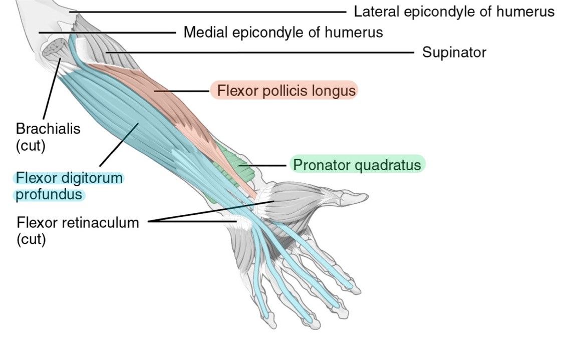
Try labeling diagrams and worksheets as additional learning aids. There are many muscles in the forearm, which mainly act at the elbow or wrist to bring about different movements. The flexor pollicis longus is situated on the radial side of the forearm, lying in the same plane as the preceding. A deep layer , intermediate layer and superficial layer. This diagram depicts muscle of the body diagrams 7441054 with parts and labels.
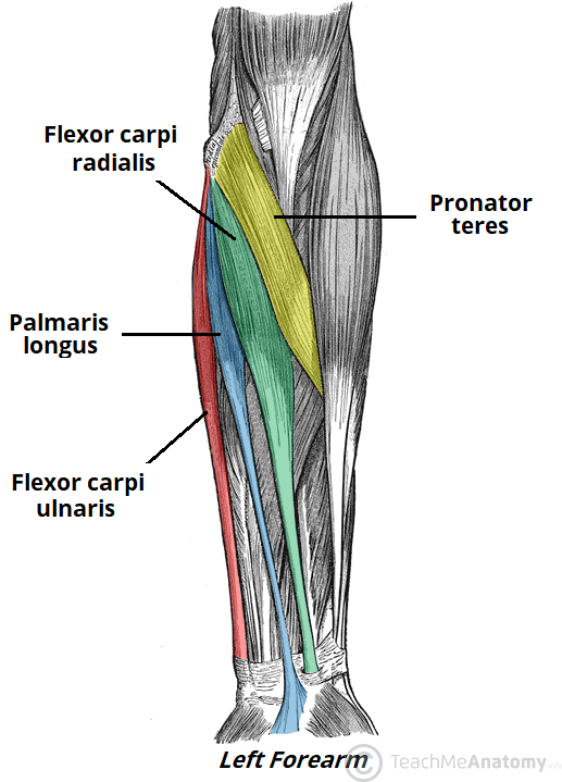
Muscles Of The Anterior Forearm Flexion Pronation Teachmeanatomy from teachmeanatomy.info I made an entire tutorial dedicated to drawing the forearms with anatomical detail, it can be fond here. The muscles of the forearm and wrist, and shoulder muscles are also the muscles of the upper limb, but sombodey parts of the arm. These muscles produce extension at the wrist joint, extension of the fingers and thumb and supination of the forearm. Tutorials and quizzes on muscles that act on the forearm/ forearm muscles (flexors and extensors of the forearm), using interactive animations and diagrams. This layer contains only one muscle, the flexor digitorum. 2, ulna, 3, biceps muscle; It has 2 heads of proximal attachment , between which the ulnar nerve passes distally in. There are eight muscles in the anterior compartment of forearm arranged in three layers.
11 photos of the forearm muscles diagram structure.
4, attachment… the muscles of the back forearm. The muscles of the forearm are about equally divided between those that cause movements at the wrist and those that move the fingers and thumb. Pronator teres pronates the forearm, turning the hand posteriorly. This layer contains only one muscle, the flexor digitorum. Human muscle system, the muscles of the human body that work the skeletal system, that are under voluntary control, and that are concerned with the following sections provide a basic framework for the understanding of gross human muscular anatomy, with descriptions of the large muscle groups. Editor · aug 11, 2017 ·. In the anterior compartment, they are split into three categories: These muscles produce extension at the wrist joint, extension of the fingers and thumb and supination of the forearm. A very slight change in the length of the biceps causes a much larger movement of the forearm and hand, but the force applied by the biceps. Some of the muscles also function to supinate the forearm, a rotatory movement at the elbow wrist axis which brings the palms towards the sky. The forearm is the region of the upper limb between the elbow and the wrist. The term forearm is used in anatomy to distinguish it from the arm. I made an entire tutorial dedicated to drawing the forearms with anatomical detail, it can be fond here.
The muscles of the upper arm are responsible for the flexion and extension of the forearm at the elbow joint. I've just switched over to a diagram to show you this muscle. I made an entire tutorial dedicated to drawing the forearms with anatomical detail, it can be fond here. Strength training exercises are common ways to increase the size and overall strength of the major muscles in the arms. The antibrachial or forearm muscles may be divided into a volar and a dorsal group.
11 Muscles Of The Forearm Simplemed Learning Medicine Simplified from simplemed.co.uk The anconeus, located in the superficial region of the posterior forearm compartment, moves the ulna during pronation and extends the forearm at the elbow. A deep layer , intermediate layer and superficial layer. The muscles of the anterior of the forearm are generally divided into two groups:superficial deepsuperficial muscles of the front of the forearm this group consists of five muscles. Pronator teres pronates the forearm, turning the hand posteriorly. Try labeling diagrams and worksheets as additional learning aids. 11 photos of the forearm muscles diagram structure. Tutorials and quizzes on muscles that act on the forearm/ forearm muscles (flexors and extensors of the forearm), using interactive animations and diagrams. The muscles of the upper arm are responsible for the flexion and extension of the forearm at the elbow joint.
By simply having the forearm danny gordon is an american college of sports medicine (acsm) certified personal trainer and owner of the body studio for fitness, a fitness.
This diagram depicts muscle of the body diagrams 7441054 with parts and labels. A deep layer , intermediate layer and superficial layer. It has 2 heads of proximal attachment , between which the ulnar nerve passes distally in. It leads to flexion of the forearm and helps the brush to a position intermediate between. It is a functionally important muscle that contains two heads. Pronator teres pronates the forearm, turning the hand posteriorly. Learn vocabulary, terms and more with flashcards, games and other study tools. Muscle diagram of shoulder human shoulder muscle other important bones in the shoulder include. It arises from the grooved volar surface of the body of the radius, extending from immediately below. I've just switched over to a diagram to show you this muscle. Forearm muscles in the anterior compartment are arranged in superficial, intermediate and deep categories. Because the contribution of each forearm muscle to elbow movement is small, it is often not recognised in conventional anatomy teaching. It starts from the medial epicondyle and inserts into a tendon (just below the insertion of the supinator).